Which skull bone contains a protuberance

Daniel Rodriguez
Published Apr 03, 2026
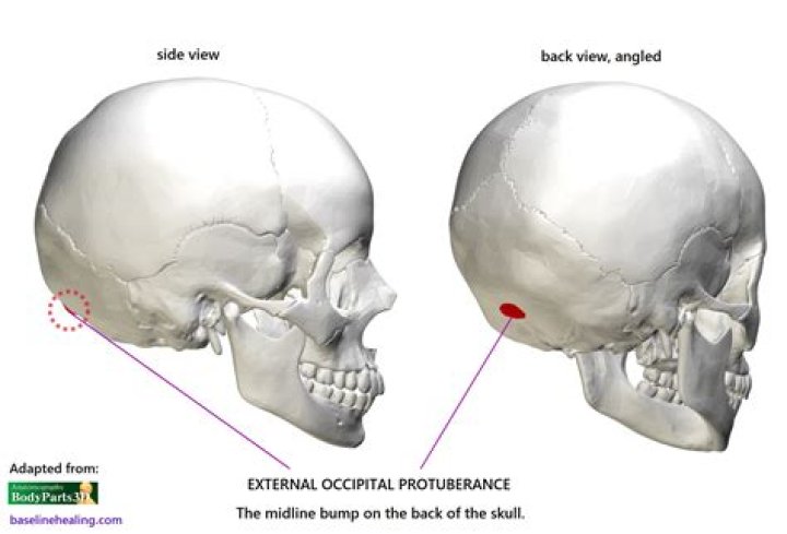
The occipital bone forms the posterior aspect of the skull and posterior floor of the cranial cavity. A prominence, the external occipital protuberance, or inion, is found on the external surface at the posterior midline (Figure 8-2). The large foramen magnum is found in the inferior aspect of the occipital bone.
Which skull bone contains a protuberance quizlet?
Terms in this set (32) The inion is the most prominent projection of the protuberance which is located at the posterior (lower rear) part of the human skull. each of two rounded knobs on the occipital bone that form a joint with the first cervical vertebra.
What bone serves as an attachment point for muscles but does not articulate with any other bone?
The primary function of the hyoid bone is to serve as an attachment structure for the tongue and for muscles in the floor of the oral cavity. It has no articulation with other bones.
Which of the following locations is not formed by part of the Maxillae?
Which of the following locations is not formed by part of the maxillae? The nasal septum is formed by the vomer and perpendicular plate of the ethmoid bone. The maxillary bone, although close in proximity, does not contribute to the structure of the nasal septum.Which bone is not associated with the skull?
The hyoid bone is an independent bone that does not contact any other bone and thus is not part of the skull (Figure 17).
Which bone does not contain a paranasal sinus quizlet?
Explanation: There are four paranasal sinuses in the head: the frontal, maxillary, sphenoid, and ethmoid sinuses. They function in lightening the skull, and creating mucous for the nasal cavity. The temporal bone does not contain a sinus.
What bones meet at the Lambdoid suture?
The lambdoid suture is a line of dense, fibrous tissue that connects the occipital bone with the parietal bones. It is continuous with the occipitomastoid suture, which connects the occipital bone with the temporal bones.
Is the ethmoid bone part of the axial skeleton?
The ethmoid bone forms the inferior portion of the nasal septum. The ethmoid bone is part of the axial skeleton.Is the hyoid bone part of the axial or appendicular skeleton?
The axial skeleton is the part of the skeleton that consists of the bones of the head and trunk of a vertebrate. In the human skeleton, it consists of 80 bones and is composed of six parts; the skull (22 bones), also the ossicles of the middle ear, the hyoid bone, the rib cage, sternum and the vertebral column.
What bones make up zygomatic arch?The cranial portion of the zygomatic arch is formed by the zygomatic bone, and the caudal portion is formed by the zygomatic process of the temporal bone. The zygomatic arch forms the ventral and lateral rim of the orbit.
Article first time published onWhich bone marking provides a site where bones articulate or to which ligaments and tendons attach quizlet?
Which bone marking provides a site where bones articulate or to which ligaments and tendons attach? projection.
Which list contains only facial bones?
which of the following lists contains only facial bones? a. mandible, maxilla, nasal, zygomatic b. frontal, occipital, zygomatic, parietal c. occipital, sphenoid, temporal, lacrimal d.frontal, parietal, occipital, sphenoidaBlood vessels that drain blood from the head pass through thejugular foramina
What cranial bone articulates with all other cranial bones?
ABThe _____ _______ of the sphenoid bone houses the pituitary gland.sella turcicaThe ________ bone is called the keystone of the cranial floor because it articulates with all other cranial bones.sphenoid
What bone is the external occipital protuberance on?
External Occipital Protuberance (EOP) is a normal anatomical structure located on the posterior surface of the occipital bone, at the level of the superior nuchal line.
Which skull bones occur in pairs?
The paired bones are the maxilla, palatine, zygomatic, nasal, lacrimal, and inferior nasal conchae bones. The unpaired bones are the vomer and mandible bones.
What bone has sella turcica?
Introduction: The sphenoid bone has a superior depression called the sella turcica, Latin for “Turkish saddle,” where the pituitary gland is found.
What are the 4 sutures of skull?
- Metopic suture. This extends from the top of the head down the middle of the forehead, toward the nose. …
- Coronal suture. This extends from ear to ear. …
- Sagittal suture. …
- Lambdoid suture.
What are the skull sutures?
The cranial sutures are fibrous joints connecting the bones of the skull. … The dense fibrous tissue that connects the sutures is made mostly out of collagen. These joints are fixed, immovable, and they have no cavity. They are also referred to as the synarthroses.
What bones meet at the coronal suture?
The coronal suture lies between the posterior border of the frontal bone and the anterior margins of the left and right parietal bones. It projects inferiorly to meet the junction of the greater wing on the sphenoid bone and the squamous part of the temporal bone.
What skull bones contain paranasal sinuses?
The large facial bones that surround the nasal cavity – the frontal bone, the maxilla, the sphenoid and ethmoid bones – are hollow to a greater or lesser extent. The hollow spaces in these bones contain the paranasal sinuses, which in the healthy living body are filled with air.
Does the frontal bone contain a sinus?
There are two, large frontal sinuses in the frontal bone, which forms the lower part of the forehead and reaches over the eye sockets and eyebrows. The frontal sinuses are lined with cells that make mucus to keep the nose from drying out. Anatomy of the paranasal sinuses (spaces between the bones around the nose).
Does the maxillary bone contain a sinus?
The maxilla is a bone which helps to make up the skull. It is specifically located in the mid face, forms the upper jaw, separates the nasal and oral cavities, and contains the maxillary sinuses (located on each side of the nose.
Is the skull part of the appendicular skeleton?
The appendicular skeleton includes all the bones that form the upper and lower limbs, and the shoulder and pelvic girdles. … The axial skeleton includes the bones that form the skull, laryngeal skeleton, vertebral column, and thoracic cage.
Where is the hyoid?
The hyoid bone (hyoid) is a small U-shaped (horseshoe-shaped) solitary bone, situated in the midline of the neck anteriorly at the base of the mandible and posteriorly at the fourth cervical vertebra. Its anatomical position is just superior to the thyroid cartilage.
Is coccyx axial or appendicular?
The axial skeleton supports the head, neck, back, and chest and thus forms the vertical axis of the body. It consists of the skull, vertebral column (including the sacrum and coccyx), and the thoracic cage, formed by the ribs and sternum. The appendicular skeleton is made up of all bones of the upper and lower limbs.
Are there two ethmoid bones?
Ethmoid boneFMA52740Anatomical terms of bone
What type of bone is ethmoid?
The ethmoid bone is an unpaired cranial bone that is a significant component of the upper nasal cavity and the nasal septum. The ethmoid bone also constitutes the medial orbit wall.
What is hyoid bone for?
Together with its attached muscles, the hyoid bone has two important functions: it holds up the tongue, which sits above it, and it holds up the larynx, which hangs below it. It also transmits the force of muscles that help to open the jaw. Let’s take a closer look at the hyoid bone.
What is zygomatic arch of skull?
zygomatic arch, bridge of bone extending from the temporal bone at the side of the head around to the maxilla (upper jawbone) in front and including the zygomatic (cheek) bone as a major portion. … The zygomatic arch is particularly large and robust in herbivorous animals, including baboons and apes.
Which bone contains the mandibular fossa?
Each mandibular fossa or glenoid fossa forms the temporal component of the TMJ. It is a concave area on the inferior border of the squamous part of the temporal bone that is also referred to as the articular fossa.
Which bone in the skull contains the auditory organs and the organs of hearing?
The temporal bone is a thick, hard bone that forms part of the side and base of the skull. This bone protects nerves and structures in the ear that control hearing and balance.