What nerves are involved in patellar reflex

Michael Henderson
Published Apr 21, 2026
The patellar tendon reflex
Which nerves are involved in reflexes?
The anatomical pathway of a reflex is called the reflex arc. It consists of an afferent (or sensory) nerve, usually one or more interneurons within the central nervous system, and an efferent (motor, secretory, or secreto-motor) nerve. Most reflexes have several synapses in the reflex arc.
Which part of nervous system controls reflexes?
Spinal cord central nervous system controls reflexes.
Which nervous system controls reflex action?
The peripheral nervous system (PNS) is a system of nerves which connect the central nervous system (CNS) (includes the brain and spinal cord) with other parts of the body. Reflex action is the result of the coordination of the spinal cord and peripheral nervous system.What nerve is tested in patellar reflex?
The patellar tendon reflex tests the function of the femoral nerve and spinal cord segments L2-L4.
What nerve carries the afferent and efferent impulses in the patellar reflex?
The quadriceps femoris reflex also called the patellar reflex, is elicited by inducing rapid stretch in the common quadriceps tendon distal to the patella (technically the patellar ligament, but in this functional context, the quadriceps femoris tendon), sending an afferent action potential to the spinal cord via the …
Which nerve is tested by tapping on the patellar ligament?
The patellar reflex, also called the knee reflex or knee-jerk, is a stretch reflex which tests the L2, L3, and L4 segments of the spinal cord.
What happens in patellar reflex?
knee-jerk reflex, also called patellar reflex, sudden kicking movement of the lower leg in response to a sharp tap on the patellar tendon, which lies just below the kneecap. … In reaction these muscles contract, and the contraction tends to straighten the leg in a kicking motion.Which nerves transmit impulses from the central nervous system?
Explanation: Motor neurons carry nerve impulses from the brain and spinal cord to muscles and glands. Interneurons carry nerve impulses back and forth between sensory and motor neurons.
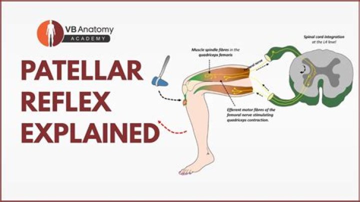
What are the steps involved in the patellar reflex?A tap to the patellar tendon stretches the quadriceps muscle (1) resulting in activation of the muscle spindle (2). The afferent neuron of the muscle spindle, detecting stretch, sends a signal to the spinal cord (3) and synapses directly with a motor neuron (4) that causes the quadriceps muscle to contract (5).
Article first time published onWhat kind of reflex is withdrawal reflex?
The withdrawal reflex is a spinal reflex intended to protect the body from damaging stimuli. It is a polysynaptic reflex, causing stimulation of sensory, association, and motor neurons.
Is patellar reflex somatic or autonomic?
Autonomic Reflexes Activity 1- Patellar reflex The patellar tendon reflex or knee-jerk reflex is a monosynaptic stretch reflex that assesses the nervous tissue between (and including) the L2 and L4 segments. It can be done by tapping the patellar ligament (just below the knee) with a reflex hammer.
Why do doctors test patellar reflex?
Medical author Dr Janice Rachel Mae explains that doctors routinely use reflex tests to check if there are any problems in the nervous system involved in movement, nerve functioning or health of the connective tissue in the knee or leg.
What type of nerve carries both afferent and efferent impulses?
Direction of Signal Transmission Efferent nerves conduct signals away from the central nervous system to target muscles and glands. Mixed nerves contain both afferent and efferent axons, and thus conduct both incoming sensory information and outgoing muscle commands in the same nerve bundle.
Which nerves carry nerve impulses away from the spinal cord quizlet?
Afferent neurons are sensory neurons that carry nerve impulses from sensory stimuli towards the central nervous system and brain, while efferent neurons are motor neurons that carry neural impulses away from the central nervous systme and towards muscles to cause movement.
Are most nerves motor nerves?
Most of the nerves have both sensory and motor components. Three of the nerves are associated with the special senses of smell, vision, hearing, and equilibrium and have only sensory fibers. Five other nerves are primarily motor in function but do have some sensory fibers for proprioception.
What causes the patellar reflex quizlet?
When patellar is stretched, the response is a rapid extension of the leg at the knee. The patellar reflux involves the rapid contraction of QUADRICEPS FEMORIS when a stretched patellar ligament is tapped. … Involves rapid contraction of the biceps brachii in response to a sudden stretch of the biceps tendon.
What muscles are involved in the knee-jerk reflex?
The knee-jerk reflex, also known as the patellar reflex, is a simple reflex that causes the contraction of the quadriceps muscle when the patellar tendon is stretched. I describe the course of the reflex arc from muscle spindles in the quadriceps muscle to motor neurons that cause movement of the leg.
What causes no patellar reflex?
[1] Many additional causes of peripheral neuropathy can yield an absent or diminished patellar tendon reflex, including diabetes, alcohol use disorder, amyloidosis, vitamin deficiencies, toxins, and remote cancer.
What is the reflex in your knee?
The reflex that the doctor checks by tapping your knee is called the patellar, or knee-jerk, reflex. It is also known as a deep tendon reflex (DTR) because the doctor is actually tapping on a tendon called the patellar (say: puh-TEL-ur) tendon.
Is the brain involved in a reflex reaction?
This quick response is called a reflex, and reflexes occur without conscious thinking or planning, meaning the brain is not involved in them.
Does the patellar reflex involve the brain?
The normal knee-jerk or, “patellar jerk,” reflex is elicited when the knee is tapped below the knee cap (patella). Sensors that detect stretching of the tendon of this area send electrical impulses back to the spinal cord. … The brain is never involved in the reflex.
What actions occur during a withdrawal reflex?
The reflex occurs when the flexors in the withdrawing limb contract and the extensors relax, while in the other limb, the opposite occurs. An example of this is when a person steps on a nail, the leg that is stepping on the nail pulls away, while the other leg takes the weight of the whole body.
What controls the withdrawal reflex?
The central nervous system is involved because the sensory neuron communicates through the spinal cord to relay the withdrawal reflex. Lastly, the muscles of the body accomplish the movement due to the reflex. Extensors and flexors complete the reflex.
What are sensory nerves?
A sensory nerve, or afferent nerve, is a general anatomic term for a nerve which contains predominantly somatic afferent nerve fibers.
Which reflexes are mediated through cranial nerves What is the purpose of this reflex?
Cranial Nerve Reflex Tests- Two reflexes mediated by cranial nerves are the corneal reflex and gag reflex. i. Corneal Reflex- The corneal reflex is mediated through the trigeminal nerve (cranial nerve V). The absence of this reflex is an ominous sign because it often indicates damage to the brain stem.
Is the knee jerk reflex innate?
Many reflexes of placental mammals appear to be innate. … Reflexes include not only such simple acts as chewing, swallowing, blinking, the knee jerk, and the scratch reflex, but also stepping, standing, and mating.