What is the function of the Hyalocytes?

Rachel Hickman
Published Feb 23, 2026
What is the function of the Hyalocytes?
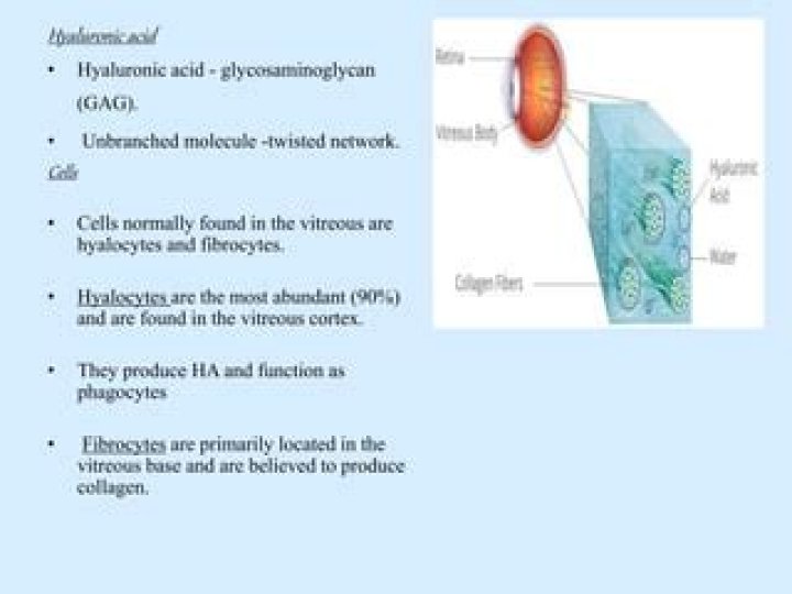
Hyalocytes, also known as vitreous cells, are cells of the vitreous body, which is the clear gel that fills the space between the lens and the retina of the eye. Hyalocytes occur in the peripheral part of the vitreous body, and may produce hyaluronic acid, collagen, fibrils, and hyaluronan.
Where are hyalocytes found?
These cells, cuurently known as hyalocytes, are located in the vitreous cavity at an average distance of 50 μm from the inner surface of the retina and are concentrated anteriorly in the vitreous base and posteriorly in the vicinity of the optic papilla.
What is primary secondary and tertiary vitreous?
Briefly, the primary vitreous is associated with the hyaloid vascular supply system, which nourishes the lens during development. The secondary vitreous is laid down around the primary vitreous and forms the definitive (adult) vitreous, whereas the tertiary vitreous contributes to the formation of the lens zonules.
What is tertiary vitreous?
The secondary vitreous is laid down around the primary vitreous and forms the definitive (adult) vitreous, whereas the tertiary vitreous contributes to the formation of the lens zonules. Figure 14-1 Vitreous after dissection of the sclera, choroid, and retina.
What is secondary vitreous?
2° vitreous –9th weeks gestation–primary vitreous degenerates to become hyaloid canal secondary vitreous develops as a relatively acellular and avascular structure. The cells of secondary vitreous are called hyalocytes.
What is ora serrata in human eye?
Anatomical terminology The ora serrata is the serrated junction between the choroid and the ciliary body. This junction marks the transition from the simple, non-photosensitive area of the ciliary body to the complex, multi-layered, photosensitive region of the retina.
What is disorder of the vitreous body of the eye?
What is vitreous degeneration? Vitreous degeneration refers to a change that occurs in the vitreous humor (or vitreous fluid) in the eye, as the vitreous humor changes from a thick vitreous gel to a thin liquid substance.
Can a vitreous detachment heal?
This is a condition where the vitreous, which was gel when the person was younger, has become liquefied and has begun to peel away from the retina. This is a natural development in the majority of people over the age of 60. It doesn’t heal, but it usually doesn’t require any treatment either.
How is aqueous humor drained?
Aqueous humour drains out of the eye through the trabecular meshwork. The trabecular meshwork is a spongy mass of tiny canals located in the drainage angle. The drainage angle is located between the iris and the clear covering of the eye (cornea), where the iris meets the white outer covering (sclera) of the eye.
What tissue is the ora serrata made of?
pigmented epithelium
This point is the ora serrata. In this region the pigmented epithelium of the retina transitions into the outer pigmented epithelium of the ciliary body and the inner portion of the retina transitions into the non-pigmented epithelium of the cilia….
| Ora serrata | |
|---|---|
| TA98 | A15.2.04.006 |
| TA2 | 6780 |
| FMA | 58600 |
| Anatomical terminology |
What are the symptoms of vitreous degeneration?
Signs and symptoms of vitreous degeneration
- A sudden increase in floaters defined as mobile blurry shadows that obscure the vision.
- Flashes or streaks of light which are most noticeable in dark surroundings.
- Seeing a curtain-like shadow coming down across the vision which is the indicative sign of retinal detachment.
How do you slow down vitreous degeneration?
There is no specific treatment for vitreous degeneration; a vitrectomy laser surgery can be performed to help alleviate any vitreous floaters.