What is internal vertebral venous plexus

John Castro
Published May 01, 2026
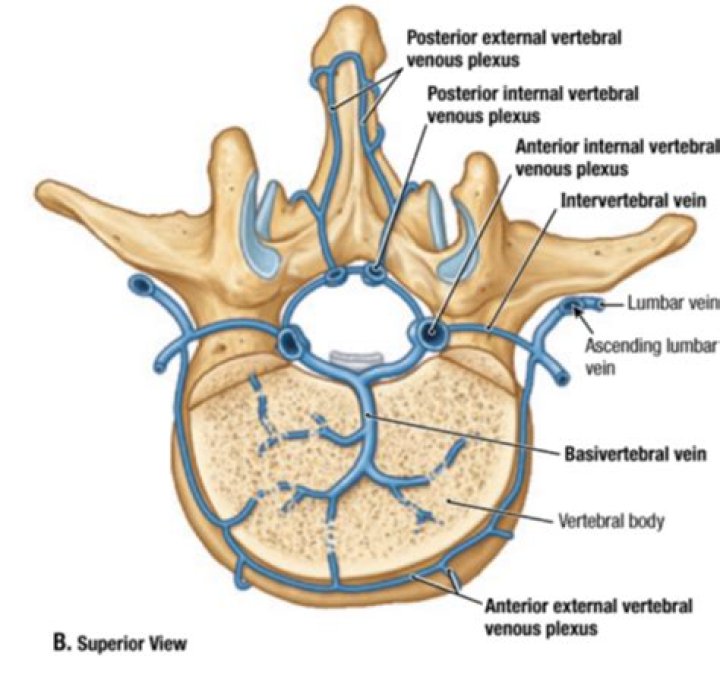
The epidural (internal vertebral) venous plexus, which drains blood not only from the cord but also from bone and red bone marrow, consists of several anterior and posterior longitudinal and interconnecting vessels. At the level of the foramen magnum, it forms a dense network that communicates with vertebral veins.
What are venous plexuses?
A venous plexus is a congregation of multiple veins.
Where does main part of vertebral venous plexus lies?
Median sagittal section of two thoracic vertebrae, showing the vertebral venous plexuses. The internal vertebral venous plexuses (intraspinal veins) lie within the vertebral canal in the epidural space, and receive tributaries from the bones and from the spinal cord.
Where does internal vertebral venous plexus drain?
The internal plexus drains into the external one, which in turn drains into the vertebral veins of the neck and segmental (intercostal, lumbar, and sacral) veins of the trunk.Why is Suboccipital venous plexus important?
The suboccipital venous plexus drains deoxygenated blood from the back of the head. It communicates with the external vertebral venous plexuses. The external vertebral venous plexuses travel inferiorly from this suboccipital region to drain into the brachiocephalic vein.
Where does prostatic venous plexus drain?
The receipt of blood from the vesical and prostatic rami connect the prostatic plexus to the vesical plexus and internal pudendal vein. The prostatic plexus then drains into the vesical and internal iliac veins.
What is a plexus?
A plexus is a bundle of intersecting nerves, blood vessels, or lymphatic vessels in the human body. These bundles typically originate from the same anatomical area and serve specific areas of the body. Bundles of nerves that form a plexus communicate information to your brain about pain, temperature, and pressure.
What is a portal venous system?
The portal venous system and hepatic veins are a paired network of valveless veins responsible for blood from all of the abdominal viscera, excluding the kidneys and adrenal glands. Before reaching the heart the blood collected by the tributaries of the portal vein passes through the hepatic sinusoids.What is the clinical significance of spinal valveless plexus?
Today, the VVP is considered part of the cerebrospinal venous system, which is regarded as a unique, large-capacitance, valveless plexiform venous network in which flow is bidirectional that plays an important role in the regulation of intracranial pressure with changes in posture and in venous outflow from the brain, …
Where is Batson venous plexus?The Batson venous plexus (Batson veins) is a network of valveless veins in the human body that connect the deep pelvic veins and thoracic veins (draining the inferior end of the urinary bladder, breast and prostate) to the internal vertebral venous plexuses.
Article first time published onWhere does the Suboccipital nerve come from?
Course: The suboccipital nerve is the dorsal ramus of the first cervical nerve. It emerges from the central canal to travel between the posterior arch of C1 inferiorly and the vertebral artery superiorly.
What is in the Suboccipital triangle?
The suboccipital triangle contains the vertebral artery, suboccipital nerve (C1), and suboccipital venous plexus. … The left and right vertebral arteries then merge at the brainstem to form the basilar artery. The suboccipital nerve is the posterior ramus of C1 spinal nerve.
Is plexus safe to take?
No serious side effects have been reported for Plexus Slim, and it appears safe overall. However, like many other diet supplements, more research is needed on its long-term effects and safety. Some people have reported unpleasant but non-serious symptoms, such as bloating, gas, nausea, stomach ache and constipation.
Where is the plexus located?
Nerve Junction Boxes: The Plexuses Four nerve plexuses are located in the trunk of the body: The cervical plexus provides nerve connections to the head, neck, and shoulder. The brachial plexus provides connections to the chest, shoulders, upper arms, forearms, and hands.
What happens if the cervical plexus is damaged?
Damage to the cervical plexus can cause sensory disturbances to the posterior head, neck, submandibular region, and the superior back, in a cape-like distribution.
What is prostatic venous plexus?
The prostatic venous plexus is a network of small veins that surrounds and drains the prostate gland of the male pelvis. It communicates with the inferior vesical vein of the urinary bladder superiorly, and the internal vertebral venous plexus posteriorly.
What is the prostatic plexus?
The prostatic plexus is a relatively large bundle of nerves that arises from the inferior (lower) portion of the pelvic plexus, a bundle of nerves, located on either side of the rectum. It is located in the prostate’s fascial shell, a layer of connective tissue.
What nerves control the prostate?
The prostate gland receives sympathetic input via the hypogastric nerve and parasympathetic input via the pelvic nerve. In addition, the hypogastric and pelvic nerves also provide sensory inputs to the gland.
What is epidural venous plexus?
The epidural venous plexus is a network of interconnecting veins located in the anterior epidural space, in the outermost part of the spinal canal. It runs from the skull base to the sacrum. It is surrounded by very little fat, although the levels increase towards the lower levels of the spine.
What does the internal jugular vein drain?
The function of the internal jugular vein is to collect blood from the skull, brain, superficial parts of the face, and the majority of the neck. … The blood collected from these vessels then drains to the brachiocephalic vein and into the right atrium.
How does the pampiniform plexus regulate temperature?
The pampiniform plexus helps regulate the temperature of the testes by acting as a “heat exchange” mechanism to cool down the blood. The arteries supplying the testes run through the plexus where the blood is cooled from abdominal arterial temperature to testicular temperature.
What is the purpose of the hepatic portal system?
The hepatic portal system is the venous system that returns blood from the digestive tract and spleen to the liver (where raw nutrients in blood are processed before the blood returns to the heart).
What is hepatic portal?
Listen to pronunciation. (heh-PA-tik POR-tul vayn) A blood vessel that carries blood to the liver from the intestines, spleen, pancreas, and gallbladder. Also called portal vein.
Does portal vein thrombosis cause pain?
Portal vein thrombosis causes upper abdominal pain, possibly accompanied by nausea and an enlarged liver and/or spleen; the abdomen may be filled with fluid (ascites). A persistent fever may result from the generalized inflammation.
What drains into pterygoid plexus?
In addition, the inferior ophthalmic vein and deep facial vein also drain into the pterygoid plexus. The plexus itself drains via the short maxillary vein before it forms the retromandibular vein. Emissary veins also anastomose between the plexus and the cavernous sinus, via the foramina ovale and lacerum.
Which veins have no valves?
The left and right superior and inferior pulmonary veins carry oxygenated blood from the lungs back to the left atrium of the heart. They differ from other veins in that they do not have valves.
What is suboccipital pain?
Pain from the suboccipital muscles commonly feels like a band wrapping around the head. Also, tension in these muscles may cause compression of a nerve that exits the base of the skull, and trigger pain that wraps over the head and above the eyes.
What are suboccipital nerves?
The suboccipital nerve (first cervical dorsal ramus) is the dorsal primary ramus of the first cervical nerve (C1). It exits the spinal cord between the skull and the first cervical vertebra, the atlas. It lies within the suboccipital triangle along with the vertebral artery, where the artery enters the foramen magnum.
How can the Suboccipital nerve be injured?
The suboccipital nerve may play a role in the pathogenesis of cervicogenic headaches and occipital neuralgia. The nerve may also be vulnerable to trauma from whiplash injuries.
What are the 8 Suboccipital muscles?
- Rectus capitis posterior major.
- Rectus capitis posterior minor.
- Obliquus capitis superior.
- Obliquus capitis inferior.
What are the triangles of the neck?
The triangles of the neck are the topographic areas of the neck bounded by the neck muscles. The sternocleidomastoid muscle divides the neck into the two major neck triangles; the anterior triangle and the posterior triangle of the neck, each of them containing a few subdivisions.