What is filopodia formation?

Emma Martin
Published Mar 13, 2026
What is filopodia formation?
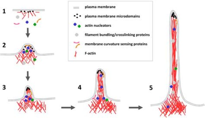
Filopodia are dynamic structures that are primarily composed of F-actin bundles and whose initiation and elongation are precisely regulated by the rate of actin filament assembly, convergence and cross-linking. Filopodia undergo 9 distinct steps in their formation.
What are podosomes in osteoclasts?
Podosomes are dynamic cell–ECM adhesive structures formed at the ventral surface of macrophages, osteoclasts and in Rous sarcoma virus-infected cells (Buccione et al., 2004; Ochoa et al., 2000).
Where are podosomes found?
plasma membrane
Podosomes are conical, actin-rich structures found on the outer surface of the plasma membrane of animal cells. Their size ranges from approximately 0.5 µm to 2.0 µm in diameter.
What are filopodia used for?
Filopodia are thin, actin-rich plasma-membrane protrusions that function as antennae for cells to probe their environment. Consequently, filopodia have an important role in cell migration, neurite outgrowth and wound healing and serve as precursors for dendritic spines in neurons.
What’s the difference between filopodia and lamellipodia?
The key difference between lamellipodia and filopodia is that the lamellipodia are cytoskeletal actin projections present in the mobile edges of the cells while filopodia are thin cytoplasmic protrusions that extend from the leading edge of the mobile cells.
How do you identify lamellipodia?
A lamellipodium borders the entire cell periphery and is punctuated by actin bundles that protrude only marginally beyond the cell edge. We have termed these bundles microspikes, to distinguish them from filopodia.
What is the difference between filopodia and lamellipodia?
Are filopodia contractile?
Filopodia formation was not inhibited and filopodia translocated into the lamella (arrowheads) but did not form contractile bundles.
Do lamellipodia use motor proteins?
Both motor proteins involve in determining the fate of lamellipodia extension by displaying distinct, but linked roles in the regulation of focal contacts formation and actin network reorganization.
What is the difference between lamellipodia and filopodia?
The key difference between lamellipodia and filopodia is that the lamellipodia are cytoskeletal actin projections present in the mobile edges of the cells while filopodia are thin cytoplasmic protrusions that extend from the leading edge of the mobile cells. Hence, they are essential structures for cell mobility.
What is marker 88?
Located on one of the few natural beaches in the Florida Keys, Marker 88 serves fresh Florida seafood, prime steaks, and chicken in an elegant Keys Casual setting on Florida bay.
What is the size of a podosome?
In particular, podosomes exhibit a short lifetime and can be found in various cell types, including macrophage, dendritic cell, smooth muscle cell, and fibroblast on viscous matrices 5, 6. Each podosome is about 0.5–2 µm in diameter and exhibits a distinct core and ring organization 7, 8.
Do podosomes exhibit mechanosensory attributes?
Adding to the known functionalities of podosomes, research suggests that these dynamic structures also exhibit mechanosensory attributes. Initial formation of podosomes seems to be influenced by the structure and composition of the underlying substratum including the presence and distribution of specific ligands.
What is the function of podosomes in melanoma?
Podosomes (yellow) in melanoma cells, along with cell nuclei (blue), actin (red), and an actin regulator (green). Podosomes are conical, actin -rich structures found on the outer surface of the plasma membrane of animal cells.