What is CD31 staining?

John Castro
Published Mar 08, 2026
What is CD31 staining?
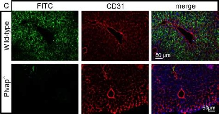
Clinical Information CD31 (cluster of differentiation 31) is expressed on endothelial cells, showing some membrane and occasional cytoplasmic staining. It is not expressed on discontinuous endothelium (eg, splenic red pulp). It is also expressed on megakaryocytes, histiocytes, plasma cells, and T-cell subsets.
What is CD31 used for?
In immunohistochemistry, CD31 is used primarily to demonstrate the presence of endothelial cells in histological tissue sections. This can help to evaluate the degree of tumor angiogenesis, which can imply a rapidly growing tumor.
What is anti CD31?
CD31 is a reported alias name for the human gene PECAM1, or ‘platelet and endothelial cell adhesion molecule 1’. The 738-amino acid protein has a reported mass of 82,522 daltons. The cellular localization is predicted to be membrane-associated. Glycosylation sites have been reported.
What is CD31 and CD34?
CD34 is one of the Endothelial Cell Differentiation Markers and Endothelial Progenitor Cell Markers. CD31 is normally found on endothelial cells, platelets, macrophages and Kupffer cells, granulocytes, T / NK cells, lymphocytes, megakaryocytes, osteoclasts, neutrophils.
What are CD31 positive cells?
CD31, also known as platelet endothelial cell adhesion molecule 1 (PECAM-1), is thought to be a sensitive and specific marker for vascular differentiation. It is a transmembrane glycoprotein expressed by endothelial cells and a variety of hematopoietic cells.
Is CD31 a surface marker?
CD31, a novel cell surface marker for CD4 cells of suppressor lineage, unaltered by state of activation. J Immunol. 1992 Jan 15;148(2):388-96.
What is if staining?
Immunofluorescence (IF) staining is a widely used technique in biological research and clinical diagnostics. IF utilizes fluorescent-labeled antibodies in order to detect specific target antigens. Followed by imaging, it is a very direct technique as you can actually see something.
What is CD34 positive?
9.1 CD34. CD34 is a transmembrane glycoprotein expressed on early lymphohematopoietic stem cells, progenitor cells, and endothelial cells. Also, embryonic fibroblasts and some cells in fetal and adult nervous tissue are CD34-positive. The TdT+ subset of precursor B cells (hematogones) is also positive for CD34.
Do all endothelial cells express CD31?
CD31 is expressed on platelets and on most leukocytes and is constitutively present on endothelial linings in vivo.
What is the difference between fluorescent and immunofluorescent?
Immunofluorescence indicates that a fluorescent tag was used to visualize the marker of interest but fluorescent markers can be used for immunocytochemistry (cells) or for immunohistochemsitry (tissues). Immunofluorescence can be used on cultured cell lines, tissue sections, or individual cells.
How is immunofluorescence performed?
Immunofluorescence (IF) or cell imaging techniques rely on the use of antibodies to label a specific target antigen with a fluorescent dye such as fluorescein isothiocyanate or cyanine dye. The primary antibody is directly conjugated to a fluorophore.
Is MSC CD34 positive?
Many studies relied on CD34 being a positive MSC marker Specifically, these investigators sorted human bone marrow nucleated cells on the basis of CD34 expression and found that greater than 95% of detectable CFU-F were recovered in the CD34+ fraction.