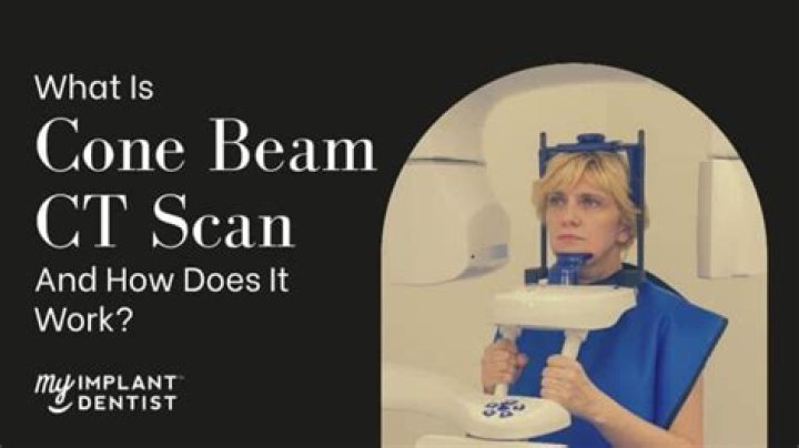
What is a cone beam xray?

Daniel Rodriguez
Published Feb 19, 2026
What is a cone beam xray?
With cone beam CT, an x-ray beam in the shape of a cone is moved around the patient to produce a large number of images, also called views. Cone beam CT provides detailed images of the bone and is performed to evaluate diseases of the jaw, dentition, bony structures of the face, nasal cavity and sinuses.
What is a cone beam image?
3D Cone Beam Imaging is an innovative x-ray scanning technology that gives your dentist the ability to see your teeth, soft tissues, and even nerve pathways with ease. This system creates a three-dimensional image of your oral cavity to give your dentist a clear and detailed view of these structures at any angle.
What is cone beam CT in Radiotherapy?
CBCT enables radiation therapists to correct for changes of the target position prior to treatment and allows monitoring of complex changes of the patient and tumor anatomy, typically caused by patient’s loss of weight and tumor regression (shape/volume changes).
What is a CBCT scan used for?
3D cone beam computed tomography (CBCT) is an imaging technology that allows dentists to evaluate the underlying bone structure, as well as the nerve pathways and surrounding soft tissues.
Are cone beam scans safe?
As outlined in the ALARA principle, every precaution should be taken to minimize radiation exposure. Radiation exposure from CBCT is up to 10 times less than that incurred from medical CT scanning, which exposes a patient to a dose of approximately 400 to 1000 µSv.
How much radiation does cone beam CT produce?
The amount of radiation received from a cone-beam CT of the jaws will vary from approximately 18–200 µSv depending on the size of the field of view, resolution of the images, size of the patient, location of the region of interest, as well as the manufacturer settings.
Is cone beam CT 3D?
Cone beam computed tomography (CBCT) is a radiographic imaging method that allows accurate, three-dimensional (3D) imaging of hard tissue structures.
What does Igrt stand for?
Image-guided radiation therapy (IGRT) is a method of radiation therapy that incorporates imaging techniques during each treatment session.
What is the difference between CT and CBCT?
A CBCT scanner uses a cone beam radiating from an X-ray source in the shape of a cone covering large volume with one single rotation about the patient. A traditional CT scanner utilizes a high-output anode X-ray tube that rotates whereas a CBCT scanner utilizes a medical fluoroscopy tube that is low-power.
Is CBCT scan same as CT scan?
CBCT is a variation on traditional computed tomography (CT) that is on the rise. Unlike traditional CT scanners, in CBCT an X-ray tube and detector panel rotate around the patient capturing data with a cone-shaped X-ray beam instead of the “slices” CT scanners are typically known for.
What is a disadvantage of CBCT?
Disadvantages of CBCT. -movement of patient causes artifacts. -size of field or view. -cost. training.
Will a CT scan show a tooth infection?
An X-ray of the aching tooth can help identify an abscess. Your dentist may also use X-rays to determine whether the infection has spread, causing abscesses in other areas. Recommend a CT scan. If the infection has spread to other areas within the neck, a CT scan may be used to assess the extent of the infection.
What is a cone beam CT system?
Cone-beam computed tomography systems (CBCT) are a variation of traditional computed tomography (CT) systems. The CBCT systems used by dental professionals rotate around the patient, capturing data using a cone-shaped X-ray beam.
What is cone beam computed tomography in dentistry?
Dental Cone-beam Computed Tomography 1 Description. Cone-beam computed tomography systems (CBCT) are a variation of traditional computed tomography (CT) systems. 2 Uses. 3 Benefits/Risks. 4 Information for Patients and Parents. 5 Information for Dental Professionals. 6 Information for Industry.
What is the difference between a cone beam and conventional X-ray?
A cone beam x-ray will provide you with a more accurate image than a conventional x-ray. A conventional x-ray only provides a 2-D image, which makes it more difficult to provide accurate treatment. A cone beam x-ray provides high-quality images, which prevents mistakes from occurring.
What is a 3-D CBCT image?
Dental CBCT images provide three-dimensional (3-D) information, rather than the two-dimensional (2-D) information provided by a conventional X-ray image. This may help with the diagnosis, treatment planning and evaluation of certain conditions.