What does Subungual melanoma look like?

Robert Miller
Published Feb 21, 2026
What does Subungual melanoma look like?
Subungual melanoma often starts as a brown or black streak under a toenail or fingernail. A person may mistake it for a bruise. Share on Pinterest A bruised nail, and dark streaks or stains on the nail with no known cause, may be signs of subungual melanoma.
Does dermoscopy detect melanoma?
Since dermatoscopes can enhance a doctor’s view of the skin, they can aid in the diagnosis of skin conditions, such as melanoma. In one 2018 review, researchers found that using a dermatoscope was more effective in diagnosing melanoma than a simple visual inspection of a skin lesion.
What does a melanoma look like under a Dermatoscope?
Characteristically, superficial melanoma is asymmetrical and irregular in shape and structure. Superficial melanomas usually have one or more of the following dermoscopic features: Blue-white veil. Multiple brown dots.
How can you tell if a nail is melanoma?
When checking your nails for melanoma, dermatologists recommend looking for the following changes:
- A dark streak.
- Dark skin next to your nail.
- Nail lifting from your fingers or toes.
- Nail splitting, which occurs when a nail splits down the middle.
- A bump or nodule under your nails.
Does nail melanoma start at the cuticle?
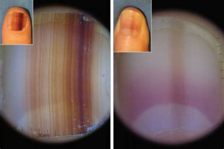
What does melanoma of the nail unit look like? Subungual melanoma often starts as a pigmented band visible the length of the nail plate (melanonychia). Over weeks to months, the pigment band: Becomes wider, especially at its proximal end (cuticle)
Does nail melanoma hurt?
In 75% to 90% of reported cases, subungual melanomas have been found in the thumb and the big toe. But they can be seen in other toes and fingers. And they can be quite painful. Inflammation, a normal bodily process that fights infection or injury, can also be present.
How accurate is dermoscopy?
The sensitivity of dermoscopy has been reported to range from 60% to 100%, depending on, among other factors, the level of experience of the examiners and the diagnostic difficulty of the evaluated lesions. Although dermoscopy improves the diagnostic accuracy for melanoma, it cannot replace histopathologic examination.
How is dermoscopy performed?
Dermoscopy is performed with a handheld instrument called a dermatoscope. The procedure allows for the visualization of subsurface skin structures in the epidermis, at the dermoepidermal junction, and in the upper dermis; these structures are usually not visible to the naked eye [2-4].
What is a Dermoscopic image?
Dermoscopy is a convenient tool to diagnose melanocytic lesions, especially nevus and melanoma. Various pigmented structures, including pigment network, dots and globules, and streaks, are observed in dermoscopy. Usually, 2D vertical images are used to explain the correlation of dermoscopy and histopathology.
What does lentigo melanoma look like?
The visual symptoms of lentigo maligna melanoma are very similar to those of lentigo maligna. Both look like a flat or slightly raised brown patch, similar to a freckle or age spot. They have a smooth surface and an irregular shape. While they’re usually a shade of brown, they can also be pink, red, or white.
How does nail melanoma start?
Subungual melanoma often starts as a pigmented band visible the length of the nail plate (melanonychia). Over weeks to months, the pigment band: Becomes wider, especially at its proximal end (cuticle) Becomes more irregular in pigmentation including light brown, dark brown.
Does nail grow back after biopsy?
It can take about 6 months to 1 year for a nail to regrow.
Can you get melanoma under your fingernails?
Other types of melanoma rarely arising under the nails are nodular melanoma and desmoplastic melanoma. Melanoma of the nail unit usually affects either a thumbnail or great toenail, but any finger or toenail may be involved. The term includes: Who gets melanoma of nail unit?
Is melanoma at nails (message) a differential diagnosis?
Melanoma at nails (message) Melanoma at nails – participate! In cases of melanonychia striata in a postpuberty patient, melanoma should be included in the differential diagnosis list.
What does dermoscopy reveal about melanoma?
In this case, dermoscopy reveals features that have been described on amelanotic melanoma of the skin. It shows an atypical vascular pattern characterized by the presence of linear and irregular vessels, the presence of milky-red areas or the presence of three or more types of vessel types within the same lesion.
Can melanoma be removed from the nail apparatus?
The melanoma must be removed surgically. This requires removal of the entire nail apparatus. Sometimes the end of the finger or toe is amputated. Some patients may be offered sentinel node biopsy to determine whether the melanoma has spread to local lymph nodes. What is the outlook for patients with melanoma affecting the nail unit?